Dermatopathology is a subject heading in pathology all unto itself. The intrinsic nature of dermatopathology specimens received in a laboratory necessitates a clear understanding of the material due to importance of the skin’s histology presentation as an organ. The goal of the histologist in the preparation of dermatopathology slides is to ensure that the entire area of skin which may contain pathology is represented in the final microscope slide.
Basic Skin Histology
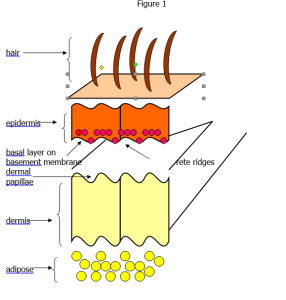
Skin anatomy and histology must be understood by histologists, and is displayed in Figure 1. Two major reasons for this are:
- The pathologist must be able to see the dermal-epidermal junction. The vast majority of skin pathology takes place in this area.
- Skin is an organ system, composed of three major tissue areas: epidermis, dermis and sub-cutaneous (adipose). This has ramifications for processing and microtomy.
Dermis / Epidermis
Skin may be thought of as being comprised of two layers: an outer epidermis and lower dermis. Adipose tissue may or may not be present below the dermis.
Notice in Figure 1 that the two layers interlock by folds in the epidermis (rete ridges) and dermis projections (dermal papillae). The epidermis sits on the basement membrane, whereon rests the basal layer of the epidermis.
The morphology (anatomy and histology) of the skin varies depending on the anatomical site. For example, hair follicles extend through the dermis into subcutis (subcutaneous fat). The skin of the face has numerous pilosebaceous elements, and the skin of the trunk has thick reticular dermis. The palms and soles have a thick compact cornified layer where usual basket-weave pattern is not seen. Muscle fibers are more prominent in the dermis of genitalia and areola. These anatomical particularities determine specifics in laboratory techniques. For example, longer fixation times and softening techniques may be necessary for specimens with a prevalence of keratotic structures.
From a technical point, the dermal / epidermal junction (DEJ) requires distinct presentation and should be clearly present in the final micro slide. Additionally,
all skin layers on a micro slide ought to be equal in morphology presentation.
Embedding
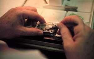
It is always a good idea to standardize procedures in the histology laboratory whenever possible. This provides the histologist with continuity in the work stream and results in increased efficiency. Embedding should be standardized in that the epidermis should be facing away from the histologist, and located at a slight angle. Additionally, the cassette back should be placed onto the mold with the number panel to the histologists left (Figures 2 and 3).

Figure 3 – The cassette back is placed onto
the mold with the number panel to the left.
Microtomy
When loading a block into the microtome for cutting, the block should be placed into the chuck with the number panel to the right. This will insure that the knife cuts from dermis to epidermis; that is from the softest tissue to the hardest. This will prevent tearing and “rolling up” of the epidermis. Sections should be cut 4 to 5 microns thick, and a very slow stroke should be used to prevent the epidermis tearing away from the dermis. Two millimeter punch specimens should be surface cut and examined unstained under the light microscope to ensure proper orientation.
Blocks of tissue that contain excess keratin and/or are very hard may be soaked in a solution of simple detergent. This is done, of course, after the block has been faced off to expose the tissue. After picking up the sections on a microscope slide and allowing them to drain, they should be placed into a conventional oven for 30-45 minutes at 60 C. After cooling, they can be stained.

The above article is based on excerpts from CM Chapman and IB Dimenstein. Dermatopathology Laboratory Techniques. Copyright 2015. In press. Please contact the author at cmchapman100@gmail.com to reserve a first print run copy of this valuable reference.
References
- Theory and Practice of Histological Techniques. Chapter 10. JD Bancroft, A Stevens ed. Churchill Livingstone, NY. Fourth edition. 1996
- Theory and Practice of Histotechnology. Chapter 9. DC Sheehan, BB Hrapchak. CV Mosby Company, St. Louis. First edition. 1980.
- Luna L. Manual of Histologic Staining Methods of the Armed Forces Institute of Pathology. 3rd ed., McGraw-Hill, New York, 1968, page 10.
- Chapman CM. Dermatopathology: A Guide for the Histologist. Copyright 2003.
- Chapman CM and Dimenstein IB. Dermatopathology Laboratory Techniques. Copyright 2015. In press.


