Histology Educational Series
Every histology laboratory has a method for preparing paraffin cell-block specimens. Certain cytology specimens, fine needle aspirates (FNAs), tiny tissue fragments, fragmented needle core biopsies and suspensions of cells for both clinical and research evaluation all require a method to get the specimen from the specimen container into a paraffin block for embedding, microtomy and staining.
Many laboratories use their own “homegrown” method (1). While these methods all vary, they have one thing in common, almost all of them produce suboptimal results in an inconsistent, non-reproducible fashion. Some of the deficiencies associated with common cell-block methods are listed below.
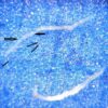
- Use of paper wraps and mesh bags/cassettes may result in the loss of cells from the specimen during processing. The pore size of the paper and the mesh size of the bags/cassettes may be larger than the diameter of a cell (i.e. average cell diameter is 50 um), resulting in the cell specimen simply washing out of the paper wrap and bags/cassettes during processing (Figure 1).
- Use of a “sponge sandwich” to hold small, friable specimens in place inside the tissue processing cassette has several deficiencies. With regard to tissue processing, the sponges may soak up and retain processing fluids during processing, resulting in contamination of processing reagents and causing suboptimal processing (2). In addition, small tissue fragments and needle biopsies may “sink” into sponges, resulting in specimen loss (Figure 2).
- Use of centrifugation and celloidin bag methods are labor intensive and still may result in specimen loss (3,4).
- Use of plasma/thrombin methods may result in brittle specimens that are difficult to section. Also, the use of pooled plasma introduces foreign DNA into the specimen, which is problematic if the specimen is to undergo additional ancillary testing for DNA.
- Some of the original specimen may be lost/discarded during the cell-block procedure. One source estimated that as many as 64% of cell-block cases were signed out as “Quantity Not Sufficient” (5).
These issues are discussed in detail in Dr. Shidham’s paper “CellBlockistry: Chemistry and art of cell-block making – A detailed review of various historical options with recent advances.” (6). Dr. Shidham defines CellBlockistry as:
“The science of exploring the chemistry and an art for achieving the best procedural outcome after processing the micro-components present in different types of specimens into the formalin-fixed paraffin-embedded (FFPE) blocks.”
As histologists, we are currently asked to provide more and more information from cell and tissue specimens that are getting smaller and smaller. This information relates to proteomic and genomic structures inside the cells, which must be preserved and processed optimally. We are also obliged to provide the entirety of the patient specimen on a slide in an optimal fashion. The loss of any of the original patient specimen is simply not acceptable.
The information contained on the microscope slide and interpreted by the pathologist is used by the clinician to not only confirm a diagnosis, but help treat the patient for a positive prognosis, so that the patient can get back to good health.
The time for Cellblockistry is now.
The next blog will describe a novel standardized cell-block procedure that provides a solution to the many deficiencies that exist in current methods that are described above.
Figure 1

Figure 2

References:
- Krogerus L. Cell Block in Cytological Diagnostics: Review of Preparatory Techniques. Acta Cytologica. 2018;62:237-243.
- Farrell DJ, Thompson PJ, Morley AR. Tissue artefacts caused by sponges.
J Clin Pathol. 1992. Oct.45(10):923-4. - Varsegi GM, Shidham V. Cell Block Preparation from Cytology Specimen with Predominance of Individually Scattered Cells. J Vis Exp. July 2009;(29):1316.
- Bussolati G. A celloidin bag for the histological preparation of cytologic material. J Clin Pathol. 1982;35:574–6.
- Krogerus L. Cell Block in Cytological Diagnostics: Review of Preparatory Techniques. Acta Cytologica. 2018;62:237-243.
- Shidham VB. CellBlockistry: Chemistry and art of cell-block making – A detailed review of various historical options with recent advances. Cytojournal. June 2019; 16:12.
- Chapman CM. CelLock: A Standardized Cell-Block Procedure. Submitted to Journal of Histotechnology; 7-11-21.
In accordance with my ethical obligation as a researcher, I am reporting that I have a financial and business interest in a company (Source Medical Products, Inc., Lake Bluff, IL) that may be affected by the research reported in the submitted paper.
Questions and comments may be sent to the author directly.
Clifford M Chapman
Senior Consultant
Medi-Sci Consultants
articles@rankinbiomed.com