Ok. I know the previous Chemistry blog may have traumatized you a bit. But hang in there – we’re almost done.
The dyes used in histology are chemical dyes that react in accordance with the basic rules of chemistry. Whether you are performing a routine hematoxylin and eosin (H&E) stain, or a special stain for fungi, the staining methods are based on underlying chemical principles.
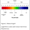
The most basic chemical principle is that of pH, which is a figure expressing the acidity or alkalinity of a solution on a logarithmic scale on which 7 is neutral, lower values are more acidic, and higher values more alkaline (basic). The “p” stands for potential and the “H” stands for hydrogen. pH is the negative log of hydrogen ion concentration in a water-based solution. Figure 1 shows the pH scale, with some examples of acidic and basic solutions. Acidic solutions have extra “H+ “molecules, and basic solutions have extra “OH- “molecules. Remember from the last blog? Rule #1 in chemistry is: opposites attract.
Basic and Acidic Dyes
The most widely used histological stains differentiate between the acidic and basic components of cells and tissues. Basic dyes have a net positive charge. They bind to components of cells and tissues that are negatively charged. Examples include: hematoxylin, phosphate groups of nucleic acids (DNA and RNA), sulfate groups of some polysaccharides (glycosaminoglycans) and some proteins (mucus)
Tissue components that stain with basic dyes are referred to as basophilic.
Acidic dyes have a net negative charge. They bind to components of cells and tissues that are positively charged. Examples include: eosin and ionized amino groups in proteins (side chains of lysine and arginine).
Tissue components that stain with acid dyes are referred to as acidophilic.
The first use of a dye is credited to Antonie van Leeuwenhoek (1673) who worked with “saffron”, a natural dye extracted from saffron crocus to stain histological structures, but genuine work on histological dye staining started not before the second half of the 19th century when C Weigher, J Gerlach, P Erlich and H Gierke systematically studied dyes for histology. At that time coloring materials were still of natural origin from which carmine or cochineal was the most used dye.
The human eye is able to perceive wavelengths of light between 400 and 700 nm. Dyes appear colored because they absorb a particular wavelength in the visible region, and the eye senses the reflected light as the complementary color.
According to their sources, coloring agents are discerned as natural or as synthetic dyes. So-called general stains will dye the tissue uniformly (indifferently) while selective stains have affinity for special cell or tissue components. A new chapter in the history of staining began when WH Perkin discovered the first aniline dye (from extracts of coal tar) in 1856 which would revolutionize the dying industry.
Dyes can be roughly divided into acidic dyes and basic dyes. Basic dyes stain acidic components, and acidic dyes stain mainly basic components. Histological staining is usually done by staining cut sections inasmuch as a dye in solution is offered to bind to defined tissue structures. Progressive and regressive techniques can be differentiated including direct and indirect procedures.
One can stain (operate) with mixtures of dyes simultaneously or successively in order to discriminate different tissue textures by the respective dyes used. So, double, triple and multiple stainings can be achieved. Many dyes have only poor affinity to tissues, but this can be overcome by the use of metal salts. Those enhancing compounds are then called “mordants”. A uniform theory of histological dye staining does not exist. This is because the mechanisms of dye binding to the different tissue components are quite heterogenous.
Hematoxylin & Eosin (H&E)
Hematoxylin and eosin, H&E or HE stain is the most commonly used technique in histology. This stain works well with a variety of fixatives and displays a broad range of cytoplasmic, nuclear, and extracellular matrix features.
Hematoxylin is a positively charged, blue dye complex. It stains basophilic structures, such as nuclei. Eosin is a negatively charged, pink dye. It stains acidophilic (also known as eosinophilic) structures. Some structures do not stain well with H&E. Hydrophobic structures tend to remain clear (such as those rich in fats).
The hallmark of an excellent H&E stain is the presence of all nuclei having been stained with precise differentiation, showing chromatin material, and nucleoli. In addition, the eosin stain should result in three shades of pink: (1) bright pink-red blood cells, (2) medium pink staining of compact connective tissue and (3) light pink staining of loose connective tissue. Figure 2 shows such and H&E stain.
If you have been able to follow the chemical principles in this blog, and the previous one, you should now have a basic understanding of the chemical principles underlying the fixation, processing and the H&E stain. Additional personal experience with regard to resolving chemistry issues in your laboratory will add to your general knowledge of chemical principles.
References
- Theory and Practice of Histological Techniques. Chapter 10. JD Bancroft, A Stevens ed. Churchill Livingstone, NY. Fourth edition. 1996
- Theory and Practice of Histotechnology. Chapter 9. DC Sheehan, BB Hrapchak. CV Mosby Company, St. Louis. First edition. 1980.
- CM Chapman. Histology Study Group. Presented at Region I meeting, hosted by MaSH. 2014.


