Hair loss and baldness in patients is referred to as “alopecia”. The hair loss can be a result of normal biology, such as male pattern baldness in men, which is a hereditary condition. Women may also experience hair loss, due to hormonal changes. There are also some medical conditions which can cause hair loss such as scalp infections (see Blog 2015 #5), thyroid imbalances, alopecia areata (i.e. auto immune attack of hair follicles), and lichen planus (inflammation caused by the immune system.
Skin specimens may be taken from the scalp in order to determine the cause of the alopecia (hair loss). These are almost always punch specimens, and are usually 3-4 mm in diameter. An obvious collection of information from such a specimen would be the number of heathy hair follicles per square millimeter. The clinician could then compare the patient result to the normal number expected, as matched for age and sex. In order to count hair follicles for comparison, these are the only skin specimens that are not cut perpendicular to the dermal-epidermal junction. Rather, they are cut parallel to it. This method was developed by Dr. Headington, and is referred to as the Headington procedure (described below).
HEADINGTON PROCEDURE FOR ALOPECIA / HAIR LOSS SPECIMENS
PRINCIPLE
Skin biopsies taken from the scalp may be for pathological diagnosis of alopecia (i.e. baldness, hair loss). It is important that the number of hair follicles can be measured. For this reason, skin specimens are prepared during the surgical gross in a particular way.
SPECIMEN
Skin specimens submitted for hair loss are almost always punch specimens.
PROCEDURE
- When the requisition is reviewed, it should be brought to the attention of the grossing technician if any of the following terms are designated: “alopecia, hair thinning, hair loss, traction, baldness”.
- If this is the case, the grossing technician will split the punch specimen , parallel to the epidermis, as follows:
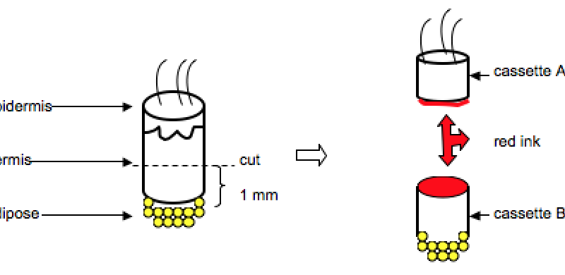
- The gross description will contain information that indicates that the “Headington” procedure was done.
- The specimens will be embedded cut surface (i.e. red ink surface) down.
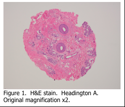
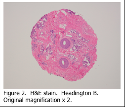
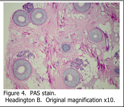
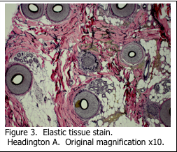
The resulting hematoxylin and eosin (H&E) slides show cross sections of the hair follicles at different skin depths (Figures 1 and 2), which can easily be counted. Additional information can be obtained by staining serial sections for elastic tissue (figure 3) and with the PAS stain for presence of fungi and/or glycogen (Figure 4). Usually, additional levels stained with H&E are also requested by the pathologist to assist with diagnosis.
REFERENCES:
- Theory and Practice of Histological Techniques. JD Bancroft, A Stevens ed. Churchill Livingstone, NY. Fourth edition. 1996
- Theory and Practice of Histotechnology. DC Sheehan, BB Hrapchak. CV Mosby Company, St. Louis. First edition. 1980.
- www.mayoclinic.org
- Dr. Terence J. Harrist – personal communication

