Anthony van Leeuwenhoek is credited with being the first person to use a microscope to view tiny “animacules” in 1674. At approximately the same time, Robert Hooke used a similar microscope to view thin slices of cork. The structures that he observed resembled the tiny cells that monks lived in at the local monastery, so he named the structures “cells”.
From that time through the 1800’s and early 1900’s, laboratorians experimented with various dyes to impart contrast to sections on microscope slides in order to see these cells. Joseph von Gerlach is credited with developing the first histology stain by using a solution of carmine in 1858 to stain brain cells. Several years later, in 1896, the hematoxylin and eosin (H&E) stain combination was worked out by Paul Mayer. The H&E stain has since remained the cornerstone of pathological diagnosis to this day.
The mid 1900’s saw the rapid development of many special stains. These stains are also used in present day histology laboratories to detect microorganisms, cell products and tissue structures. Similar to the H&E stain, these special stains use colored dye solutions to impart color and contrast to the desired structures. However, many times cells under study appear normal with these stains, even though they are later proved by other methods to display pathology.
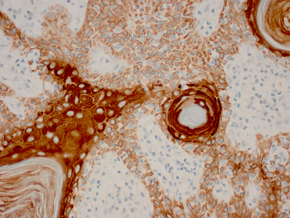
In the 1960’s and 1970’s a novel method was developed which helped pathologists to diagnose cancer cells. The method was based on using antibodies to specifically bind to protein antigens within the cells and tissue elements of a tissue section on a microscope slide. This was followed by using a “detection chemistry” to visualize the binding, such that it could be seen using a light microscope. The method is called immunohistochemistry, or “IHC” for short.
Since that time many thousands of antibodies have been developed for use in IHC. Each one will specifically bind with only one protein site. Through experimentation and research, pathologists now have a large menu of different proteins which can be detected and visualized within the specimens on the microscope slides. The presence or absence of these proteins provides information from which to form a diagnosis.
The IHC method relies on the ability to develop and use antibodies that are directed to, and bind only with, very specific proteins. Early literature described the binding as a “lock and key” arrangement. However, it was soon discovered that the binding of an antibody to its protein antigen counterpart is a very complex, three-dimensional event.
Mammals have an immune system that makes antibodies (Ab) to foreign proteins, for example, bacteria and viruses. If you inject mice, rabbits, or other animal subjects with pure protein preparations, their immune system will make antibodies to the protein. These proteins are also referred to as antigens (Ag). The antibodies are highly specific – each will bind to only one antigen.
For example, if you are exposed to the chicken pox virus, you most likely will contract the chicken pox disease. For 2-4 weeks from the exposure time, your immune system makes antibodies to the chicken pox virus. From that point on, any time you are exposed to the chicken pox virus, the antibodies circulating in your blood protect you from the disease by binding with and inactivating the chicken pox virus particles.
In histology, we can use pure antibody preparations to specifically bind to protein antigens in tissue sections. Since antibodies themselves are too small to be seen under a microscope, we can “label” them with colored tags which can be observed under the microscope. The proteins can be localized within cells, outside of cells, or even within cell nuclei.
Future segments of the educational series will delve into the methodology of this fascinating technique.

References:
1. Chapman C.M. (2017). The Histology Handbook. Amazon CreateSpace Independent Publishing Platform
2. Chapman C.M., Dimenstein I.B. (2016). Dermatopathology Laboratory Techniques. Amazon CreateSpace Independent Publishing Platform
3. https://www.nature.com/milestones/milelight/full/milelight02.html
4. Von Gerlach, J. (1858). Mikroskopische Studien aus dem Gebiet der menschlichen Morphologie (Enke)
5. Sternberger, L.A., Hardy, P.H. Jr, Cuculis, J.J. & Meyer, H. G (1970). The unlabeled antibody enzyme method of immunohistochemistry: preparation and properties of soluble antigen-antibody complex (horseradish peroxidase-antihorseradish peroxidase) and its use in identification of spirochetes. J. Histochem. Cytochem. 18, 315–333


