Every histology laboratory must have a slide labeling system. If you work in a small laboratory, this system may be as simple as using a solvent-resistant pen to hand-write your slides. Larger laboratories have moved forward to incorporate bar-code tracking systems into their LIS that can integrate with a slide printing module.
Studies have shown that handwriting slides in a histology lab can result in an error rate of 0.1 to 3%, while implementation of an automated slide printing system can reduce this rate to virtually zero (1). If you work in or supervise a histology laboratory that uses a manual slide writing system, it’s time to consider the use of a slide labeling system.
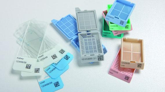
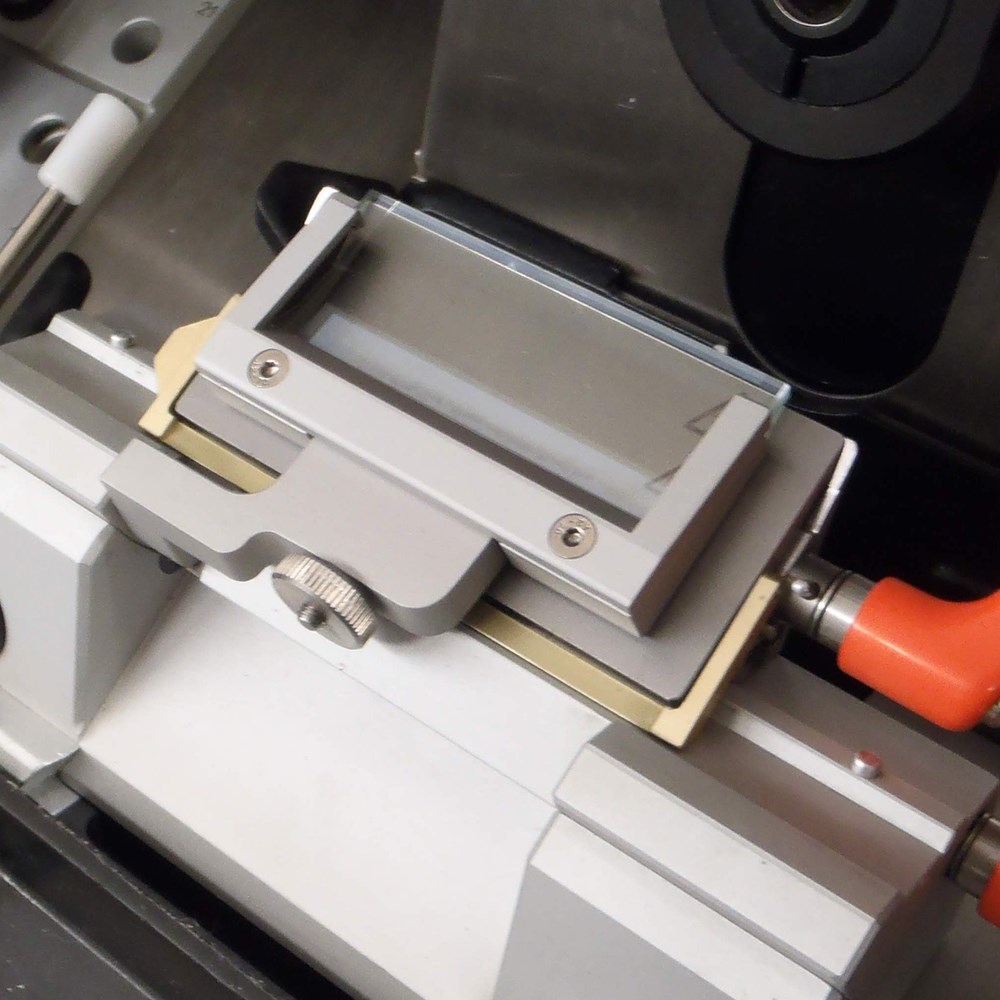
Slide labeling hardware exists in two formats. Once the system prints chemical-resistant labels that are printed, on demand by the microtomist, one case at a time. The second system prints/etches the information directly onto the microscope slide, again on demand by the microtomist, one case at a time. This procedure must be followed, instead of printing a “batch” of slides, to ensure that the microtomist is not “thumbing through” a stack of slides, looking to match the case. Any time a human is looking at a block or slide to match provides an opportunity for error.
Both CLIA and CAP regulations are clear with regard to the information that is required to be contained on a slide label. The following is found in the CLIA regulations (2)
CFR 42, IV (g) Part 493:
§ 493.1219 Condition: Histopathology.
If the laboratory provides services in the subspecialty of Histopathology, the laboratory must meet the requirements specified in §§ 493.1230 through 493.1256, § 493.1273, and §§ 493.1281 through 493.1299.
493:1291 The test report must indicate the following:
- For positive patient identification, either the patient’s name and identification number or a unique patient identifier and identification number.
CAP NSH Guidelines for “Uniform Labeling of Blocks and Slides in Surgical Pathology” (3) include the following that are pertinent to slide labeling:
- Laboratories should ensure that all blocks and slides are clearly labeled using two patient identifiers.
- Laboratories should ensure that the accession designation used on the surgical pathology report and all blocks and slides from that accession include the case type, year, and unique accession number.
We can see that CAP and CLIA agree: each slide and block must have printed on it two unique patient identifiers. Commonly this would be the patient name and accession number.
The CAP NSH Guidelines continue with the following related guidelines:
- Laboratories should label each specimen container with a unique alpha-numeric designation that incorporates the accession designation. Each block and slide from that specimen container should be labeled with the same unique alpha-numeric designation.
- When multiple slides are cut from a single block, laboratories should label each slide sequentially in order of cutting.
- The laboratory should label the slides with the histochemical, immunohistochemical, and/or special procedure.
- On microscopic slides, the accession designation should be the most prominent printed element (i.e. larger font or bolded) followed by the patient name or other second identifier and stain/procedure name. As long as the ability to read these essential elements is not compromised, additional elements may be included as determined by the laboratory.
This last guideline gives us an example of the total information required on a slide label. It is difficult, indeed, to hand-write all of this information on every single slide that is being processed in your laboratory; and to do it accurately. As referenced above, the error rate for handwriting slides ranges from 0.1 to 3%. An additional study found that 35% of the errors made in a hospital histology laboratory were made during the manual labeling of microscope slides (4).
There are additional factors that support the use of automated slide labeling in the histology laboratory. One of these is: time. It takes laboratory personnel much time, effort and concentration to label microscope slides. These same laboratory personnel could be doing other histology tasks (i.e. embedding, microtomy, slide staining, etc.). Currently, histology laboratories are being asked to run more efficiently, given the continuing decreases in laboratory services reimbursement. The decreases for reimbursement of CPT codes in pathology average minus 5% (5). Simply put: histology labor costs money – and time is money. Automating slide labeling allows personnel to be reassigned to other tasks, thereby increasing laboratory efficiency.
Also, there is the issue of risk. As mentioned above: a laboratory that goes from manual slide labeling to automated slide labeling experiences a decrease in error rate from an average of 2% to zero. That means that the risk of a misdiagnosis, whose root cause was in slide labeling, is now gone; it’s zero. Your laboratory management team and/or risk assessment officer can tell you how much it would cost for your facility to experience just one medical-legal misdiagnosis case. One source stated:
“What is the cost savings of a tracking system? It’s not an issue of using fewer reagents; it’s the staff hours saved,” … “However, the principles and the cost-benefit analysis for a tracking system would be the same regardless of your size, in my opinion. One mistake, one harmed patient, and one lawsuit can cost millions of dollars.” (1)
The final reason for using an automated slide labeling system is simple: it increases patient safety. We all want to provide our patients with a diagnosis that is 100% accurate. A slide labeling system is a tool to help us do that in our histology laboratories.
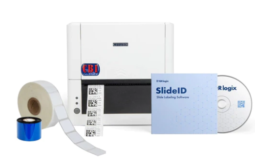
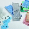
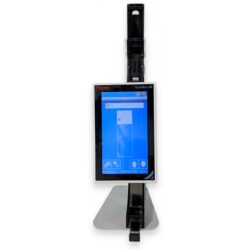
QRlogix SlideID Printer Package, stick-on slide labeling
SlideID is the only turn-key, ready-to-print microscope slide label printing solution available.
PC-based SlideID software allows you to input sample jar and slide data, stain type, and other critical details through keyboard entry. Then print the sequentially numbered slides with the click of a button.
- Arrives READY-TO-PRINT
- Keyboard or barcode scanner data entry
- On-demand printing or batch printing
- Prints sequentially numbered slides
- Optional barcode scanner – pre-programmed
- Optional PC or Tablet with pre-installed software setup
- Supports laboratory best practices
References:
- A Paxton. CAP Today. AP Tracking: an eagle eye on blocks and slides. Feature Story. February 2013.
- CLIA regulations. https://www.ecfr.gov/current/title-42/chapter-IV/subchapter-G/part-493
- CAP NSH. Guidelines for Uniform Labeling of Blocks and Slides in Surgical Pathology” March 2021. https://www.cap.org/protocols-and-guidelines/cap-guidelines/current-cap-guidelines/uniform-labeling-of-blocks-and-slides-in-surgical-pathology
- P. Morelli, E. Porazzi, M. Ruspini, U. Restelli and G. Banfi, Analysis of errors in histology by root cause analysis: a pilot study, Journal of Preventive Medicine and Hygiene, 2013 Jun; 54(2): 90–96
- 2023 Medicare PFS: See Impact to Labs and Pathology https://www.lighthouselabservices.com/2023-medicare-pfs-see-impact-to-labs-and-pathology/ogy
- Chapman CM. The Histology Handbook. Amazon CreateSpace Independent Publishing Platform; 2017.
- Chapman CM, Dimenstein IB. Dermatopathology Laboratory Techniques. Amazon CreateSpace Independent Publishing Platform; 2016.
Questions and comments may be sent to the author directly.
Clifford M Chapman | Senior Consultant
Medi-Sci Consultants
Histology Educational Series – Part 1: Slide Labeling

Rankin Biomedical moves headquarters to 35,000 square-foot facility
The Anti-Roll Plate Issue: Preventing Section Bunching
CelLock TM: An innovative standardized cell-block preparation procedure
CellBlockistry – The Time is Now